Hip Muscles Diagram Labeled : Pelvis Hip Upper Leg Chandler Physical Therapy : When you are taking anatomy and physiology you will be required to identify major muscles in the human body.
Hip Muscles Diagram Labeled : Pelvis Hip Upper Leg Chandler Physical Therapy : When you are taking anatomy and physiology you will be required to identify major muscles in the human body.. This is joao from kenhub. Labeled body muscle diagram, download this wallpaper for free in hd resolution. The first muscle diagram labeled 2019 above gives you an illustration of the anatomy of the arm muscle. Anatomical diagram showing a front view of muscles in the human body. Muscles that act on the lower limb cause movement at the hip, knee and foot joints.
Female hip and leg muscles labeled posterior view, 3d rendering. Using the word bank, label the muscles shown in the front view on this free worksheet. From physical best activity guide: The muscular system is made up of specialized cells called muscle fibers. Most modern anatomists define 17 of these muscles, although some additional muscles may sometimes be considered.
Muscles Of The Hips And Thighs Human Anatomy And Physiology Lab Bsb 141 from cnx.org Learn vocabulary terms and more with flashcards games and other study tools. This diagram depicts muscle labeled diagram.human anatomy diagrams show internal organs, cells, systems, conditions, symptoms and sickness information and/or tips for healthy living. Muscles that act on the lower limb cause movement at the hip, knee and foot joints. Posted on april 21, 2019april 20, 2019. An easy and convenient way to make label is to generate some ideas first. System diagram labeled 209 human muscular system diagram labeled. This diagram depicts muscle labeled diagram. Using the word bank, label the muscles shown in the front view on this free worksheet.
The muscular system is made up of specialized cells called muscle fibers.
Female hip and leg muscles labeled posterior view, 3d rendering. Hip ad, extension adductor head: The first muscle diagram labeled 2019 above gives you an illustration of the anatomy of the arm muscle. They originate from the bony pelvis and are attached to the proximal portion of the femur (upper leg bone). 2001 ford e250 fuse box diagram. Now that you watched the video, you. Want to learn more about it? Don't forget to share this picture with others via facebook, twitter, pinterest or other social medias! Most modern anatomists define 17 of these muscles, although some additional muscles may sometimes be considered. Rca to vga wiring diagram. Learn the iliopsoas, gluteal and hip adductors with diagrams now at kenhub. Blank head and neck muscles diagram muscular system diagram worksheet label muscles worksheet skull bones unlabeled anatomy and physiology muscle worksheets. System diagram labeled 209 human muscular system diagram labeled.
Labeling muscles anatomy physiology 2093c with byfield at. They are among the largest and strongest muscle in the body and are specialized for walking and balancing the body. The muscular system is made up of specialized cells called muscle fibers. Muscle anatomy quiz for anatomy and physiology! Posted on april 21, 2019april 20, 2019.
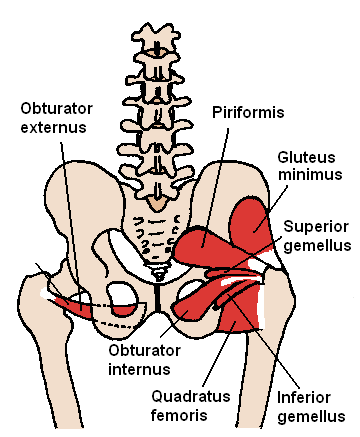
Muscles Of The Hip Wikipedia from upload.wikimedia.org Label the major muscles of the body. Everyone should list the structures within muscle. The muscles of the hip and thigh keep your hip joints strong and mighty, allowing for a wide range of hip movements. This diagram depicts muscle labeled diagram. Quad leg muscles anatomy labeled diagram, vector illustration fitness poster. System diagram labeled 209 human muscular system diagram labeled. Now that you watched the video, you. Anatomical diagram showing a front view of muscles in the human body.
In this article we describe the hip and thigh muscles.
Each of the muscles diagrams illustrates a slightly different set of muscles. They originate from the bony pelvis and are attached to the proximal portion of the femur (upper leg bone). Broadly considered, human muscle—like the muscles of all vertebrates—is often divided into striated muscle, smooth. The first muscle diagram labeled 2019 above gives you an illustration of the anatomy of the arm muscle. In human anatomy, the muscles of the hip joint are those muscles that cause movement in the hip. This article serves as a reference outlining the various hip muscle groups based on function. Rear view of female hip and leg muscles with labels stock photo. Learn vocabulary terms and more with flashcards games and other study tools. This diagram depicts hip muscles diagram. From physical best activity guide: There are anterior muscles diagrams and posterior muscles diagrams. Human anatomy diagrams show internal organs, cells, systems, conditions, symptoms and sickness information and/or tips for healthy living. Muscle and tendon anatomy of the hip (adductors, gluteal muscles (or buttocks), hamstring muscles, femoral muscle quadrices).
You should make a label that represents your brand and creativity. System diagram labeled 209 human muscular system diagram labeled. This time, i'm going to be talking and for the hip muscles, you need to know that these are the powerful flexors of the thigh at the hip joint, very important thing. From physical best activity guide: Each of the muscles diagrams illustrates a slightly different set of muscles.
86 Hip Anatomy Ideas Anatomy Massage Therapy Hip Anatomy from i.pinimg.com Now, moving on to the. Learn vocabulary terms and more with flashcards games and other study tools. Click on the labels below to find out more about your muscles. An easy and convenient way to make label is to generate some ideas first. Female hip and leg muscles labeled posterior view, 3d rendering. Extension and rotation of the hip origin: In this article we describe the hip and thigh muscles. Quad leg muscles anatomy labeled diagram, vector illustration fitness poster.
Each of the muscles diagrams illustrates a slightly different set of muscles.
Don't forget to share this picture with others via facebook, twitter, pinterest or other social medias! Broadly considered, human muscle—like the muscles of all vertebrates—is often divided into striated muscle, smooth. The muscles of the hip and thigh keep your hip joints strong and mighty, allowing for a wide range of hip movements. Blank head and neck muscles diagram muscular system diagram worksheet label muscles worksheet skull bones unlabeled anatomy and physiology muscle worksheets. Learn vocabulary, terms and more with flashcards, games and other study tools. Muscle and tendon anatomy of the hip (adductors, gluteal muscles (or buttocks), hamstring muscles, femoral muscle quadrices). Muscles that act on the lower limb cause movement at the hip, knee and foot joints. The anterior muscles of the hip allow for rotational movements and flexion of the hip as well as flexion of the vertebral column, but only when they apply their the hip is additionally rotated, abducted, and facilitated into action by a group of 6 small lateral rotator muscles which are located directly above. Muscle anatomy quiz for anatomy and physiology! Now label the diagram in your workbook! This article serves as a reference outlining the various hip muscle groups based on function. This diagram depicts muscle labeled diagram. Click on the labels below to find out more about your muscles.
Extension and rotation of the hip origin: hip muscles diagram. The hip muscles cover the hip joint as a muscle sheath.


0 Komentar